Navicular is one of those words that can make even the toughest horse owner’s heart leap. For years it has also spelled the end for a horse’s career.
Fortunately, while navicular disease can certainly complicate your riding goals, these days the diagnosis isn’t nearly as dismal.
In recent years, vets have moved away from the term navicular syndrome, as it’s not specific enough to describe what’s often going on in a case of lower-limb lameness. Now, you’re more likely to hear heel pain or caudal heel syndrome until the root cause can be narrowed down.
Navicular syndrome is classified by its degenerative impact to the navicular bone itself. Again, too specific to describe the many number of reasons for lameness in the navicular area. There can be pathology in the navicular bursa, just below the navicular bone or soft tissue around the area such as the deep digital flexor tendon and the articulation point of the coffin, pastern, and navicular bones for example.

The New and Old of Diagnosis
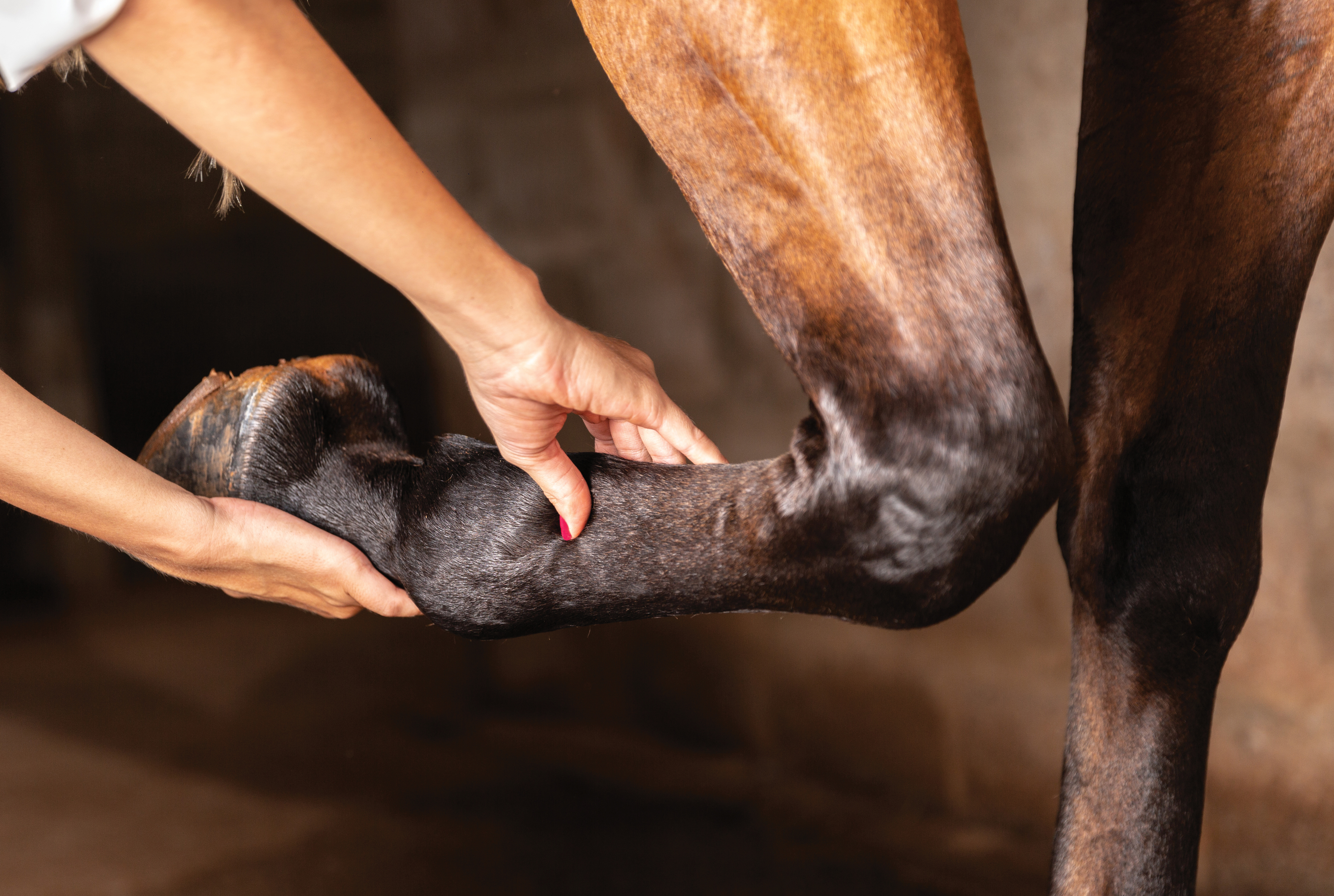
There are a few approaches to pinpoint the area of concern, which will likely start with a general soundness exam. This allows a vet to see your horse move on hard and soft ground and gather information about his symptoms. Hoof testers are also typical of early diagnosis as almost all horses with navicular-area lameness will show sensitivity over the frog and heel.
Imaging, including X-rays, MRIs, and ultrasound previously have been common for diagnosis. However, the results can be inconclusive. It’s not uncommon for an unsound horse to have clean imaging results and a perfectly sound horse to show irregularities. Imaging can be a helpful tool though so shouldn’t be thrown out; it just shouldn’t be the only approach.
Nerve blocks have become almost standard as diagnostic approaches. A palmar digital nerve block can show the extent of the lameness, with the vet applying local anesthetic to the nerve of the lower pastern area in one leg. You’ll be able to see the difference between the sound and blocked leg due to the lack of sensation—this is assuming that unsoundness is experienced in two legs, which is most common. A peri-neural or intra-articular anesthesia also may be applied locally. Your vet will work their way up your horse’s leg, and when your horse trots off soundly, you’ve found the culprit, or at least the region of concern.
Therapeutics for Repair and Management
Recovery or maintenance starts at the feet. Medication and other treatments won’t be as effective if your horse’s feet aren’t as mechanically sound as possible. Corrective shoeing might be required, which can include fixing long toes and retracted heels, and allowing the foot to expand. A wedged shoe may alleviate some tension to the deep digital flexor tendon, if that’s the issue, or a slight rocker shoe can provide some support with breakover if your horse is leading or stabbing with his toe. Your farrier should work with your horse’s foot, using radiographs for analysis if needed.
Short- and long-term pain can be mitigated with phenylbutazone (Bute) and flunixin meglumine (Banamine). Equioxx, released a few years ago, is an alternative NSAID for horses with osteo-degeneration. It’s a useful alternative, as it doesn’t seem to cause the same gastrointestinal issues of long-term Bute and Banamine use.
Finally, there are some experimental surgeries that could offer relief, but they’re far from considered best practice.

A New Solution
Biphosphonates are a relatively newer treatment option for horses suspected to have navicular syndrome, or degeneration in the bone. Osphos works by stunting the bone regeneration process, specifically slowing the action of osteoclasts, or cells that break down bone, and is given as an intramuscular injection. If degeneration is the issue, medications can slow the process, but it won’t reverse it. Your horse will remain rideable as long as the condition hasn’t progressed to the point pain is unmanageable.


